Years are no longer urging people to be "small". How to build a bone wall?
Years are no longer urging people to be "small". How to build a bone wall? May 21, 2019 Source: Voice of the Chinese Academy of Sciences - "How tall are you, grandfather?" - "There is only one meter six now. I want to be a year old. When I was young, I also had one meter seven, but now I am old, my back is also a camel, and people are short." I believe that many people have had such a dialogue with their elders, but why do people become old and dwarf like "shrinking"? The answer is - osteoporosis. How to build a "wall" of bones? Osteoporosis refers to the appearance of many voids in the bones, the density and quality of the bones also decreasing, and the bones become porous and fragile. Osteoporosis occurs in postmenopausal women and older men, with more than 200 million osteoporosis patients worldwide. One third of women over the age of 50 and one in five men are suffering from osteoporosis. In patients with osteoporosis, due to the appearance of bone voids, the density of bones is getting smaller and smaller, and the supporting force of bones is worse, which causes patients to have short height, hunchback and other symptoms, and is prone to fracture. The development and formation of bones is coordinated by osteoclasts and osteoblasts, which function functionally. Osteoclasts mainly remove old bone cells of senescence and necrosis by bone resorption; osteoblasts are responsible for secreting bone matrix after the old bone cells are removed, and the bone matrix mineralizes to form new bone. The balance between osteoclast and osteogenesis is the key to maintaining normal bone mass. We can compare the bone to a wall made up of countless bricks, and the cells that make up the bones are like bricks in the wall. When the bricks age, the osteoclasts remove the bricks, and the osteoblasts place the new bricks in the removed position for repair. In osteoporosis patients, osteoblasts and osteoclasts are unbalanced, bone resorption increases, and bone formation decreases. Equivalent to osteoclast removal bricks faster than osteoblast repair rate, in the long run, there will be a lot of holes in the wall, and finally can not support the body of this big house, thus showing a series of symptoms. PTH1R: therapeutic target for osteoporosis Parathyroid hormone (PTH) is a typical endocrine hormone that was identified as a key factor in regulating blood calcium levels more than 80 years ago and is essential for maintaining ionic homeostasis and bone health. PTH specifically binds to the type 1 parathyroid hormone receptor (PTH1R), which is highly expressed in bone cells and kidney cells, and activates downstream signaling pathways, thereby regulating calcium and phosphorus metabolism in the body. Therefore, PTH1R is a recognized therapeutic target for osteoporosis. Currently, related drugs (PTH analogs such as teriparatide) have been used in clinical practice, but the drug is a polypeptide, which can only be injected like insulin, and the treatment cost is high. . Although scientists around the world have made a lot of efforts to develop oral drugs for the treatment of osteoporosis, there are still no effective oral drugs, in part because of the lack of detailed structural information on the interaction between PTH and PTH1R. A few days ago, the Shanghai Institute of Materia Medica Xu Huaqiang and the Wang Mingwei team, the Zhejiang University School of Basic Medicine Zhang Yan team and the University of Pittsburgh School of Medicine Jean-Pierre Vilardaga team applied cryo-electron microscopy technology, accurate (resolution = 3.0Ã…) analysis of PTH1R The three-dimensional structure of the complex with Gs protein. This structure shows that the complex of long-acting activated parathyroid hormone LA-PTH with PTH1R and Gs protein consists of three layers, the first layer is the extracellular region of the receptor, and the second layer is surrounded by dish-shaped micelles. The transmembrane region of the receptor, the bottom layer is the Gs protein trimer. Among them, LA-PTH is anchored in the extracellular region and transmembrane region of the receptor in a continuous single helix form. The interaction of LA-PTH with the transmembrane region accounts for more than 60% of the interaction of the embedded region. This indicates that the interaction of this region provides the primary binding force for complex formation. Furthermore, in the complex, the extracellular domain has three different conformations, while the transmembrane region maintains the same conformation at all times. The first two conformations are similar, and the extracellular region and LA-PTH are surrounded by each other at a 15 degree angle. In the third conformation, LA-PTH is curved, which weakens the interaction with the extracellular region, suggesting LA- The interaction of PTH with the transmembrane core region provides the primary energy for receptor activation, while the extracellular region is required for binding of the promoter polypeptide to the receptor. The analysis of this structure provides an important reference for a deeper understanding of the mechanism of action of PTH-activated receptors and downstream Gs proteins. The structure is docked with the structure of the small molecule compound to virtually screen the small molecule ligand of PTH1R. That is to say, the receptor structure acts as a lock, and the small molecule compound acts as a key. If the structure of the two matches exactly, the compound can open the lock of PTH1R, which is our candidate compound. Candidate compounds obtained through virtual screening further develop therapeutically promising drugs through high-throughput screening, functional verification and structural engineering at biochemical and/or cellular levels. The work was published in the journal Science in April 2019, DOI: 10.1126/science.aav7942. The article links to https://science.sciencemag.org/content/364/6436/148.long.
Food-derived peptide is a new type of protein hydrolyzed product made from edible protein through enzymatic hydrolysis, separation and purification. Food-derived peptides come from a wide range of sources. According to the source of protein raw materials, they can be divided into animal-derived protein peptides and plant-derived protein peptides. Food-derived peptides come from a wide range of sources. According to the source of protein raw materials, they can be divided into animal-derived protein peptides and plant-derived protein peptides [1] . Food-derived peptides are divided into food-derived oligopeptides (molecular weight less than 1 000 Da) and food-derived polypeptides (molecular weight greater than 1 000 Da) according to their relative molecular mass distribution (referred to as molecular weight). Animal protein peptides are more researched on milk protein peptides, marine fish protein peptides, collagen peptides and egg white protein peptides; plant protein peptides are more researched on soybean peptides, corn peptides, wheat peptides, etc.we have Orange Juice Powder,Apple Juice Powder,Mulberry Juice Powder,Blackberry Juice Powder,Seabuckthom Juice Powder,organic garlic powder,organic sea-buckthorn fruit powder,Pomegranate Juice Powder.
Food peptide,Nutrition Supplements,Food Supplement,Amino acid Xi'an Henrikang Biotech Co.,Ltd , https://www.xianhenrikangbio.com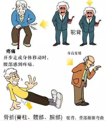
Figure 1 Symptoms of osteoporosis patients (picture from the network) 
Figure 2 Normal human bone (left) and osteoporosis patient bone (right) control (image from the network) 
Figure 3 The basic mechanism of osteoporosis (image source from the network) 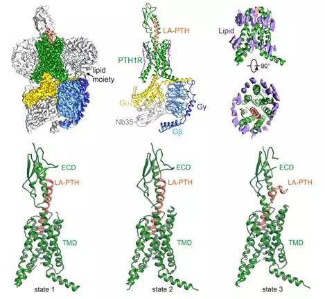
Figure 4 is a cryo-electron microscopic structure of the LA-PTH-PTH1R-Gs protein trimer complex.