[Inventory] Nature Methods: The eight most noteworthy medical technologies in 2016
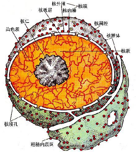
The first issue of the medical journal "Nature Methods" in the first year of the 2015 technology, selected the most concerned technical achievements: single-particle cryo-electron microscopy (cryo-EM) technology. In addition, the magazine also compiled the eight technologies that are most worthy of attention in 2016, namely: Protein labeling in cells, Unraveling nuclear architecture, and Protein structure through time. ) Precision optogenetics, Highly multiplexed imaging, Deep learning, Subcellular maps, and Integrated single-cell profiles. New protein labeling Fluorescent chemical dyes are relatively small with good photophysical properties and spectral spans. These properties make fluorescent dyes particularly attractive and are expected to replace fluorescent proteins for protein labeling. Researchers are actively developing tools to label proteins of interest in living cells with dyes. For most applications, fluorescent dyes need to be able to achieve specific labeling. There are already tools available to do this, such as the SNAP and Halo tags, the FlAsH and ReAsH, and the hexahistidine tags. These tools primarily target small proteins or polypeptides that specifically bind to the corresponding dye. Another method is to incorporate unnatural amino acids into the protein translation process. These unnatural amino acids fluoresce themselves or can be fluoresced by click chemistry. Although these methods are becoming more and more popular, they also encounter some problems. For example, fluorescent dyes have limited utility in multiple imaging, and dyes that cross the living cell membrane are less efficient, less numerous, and of lower quality. The development of such dyes is currently a fairly active area of ​​research. There is no doubt that in the future people will greatly enhance the marking efficiency of fluorescent dyes, which will further enhance the ability of quantitative imaging. Significant improvements can be made with available dyes, which increases multiplex and reduces the light required for imaging. New protein labeling methods will also appear in front of people. Specific protein labeling facilitates ultra-high resolution imaging in both fixed and living cells. When the resolution is close to tens of nanometers, the problem caused by the mark will be highlighted. For example, labeling with an antibody increases the target structure by about 10 nm, while the secondary antibody further increases the detection target. To solve this problem, many researchers are developing nanobodies. Nanobodies are small antibody fragments derived from camels and are single-domain antibodies obtained by genetic engineering methods for cloning the variable region of camelid heavy chain antibodies. Compared with traditional IgG antibodies, Nanobodies have the characteristics of small molecular mass, easy production, good stability and high antigen binding ability. We believe that more and better protein labeling strategies will emerge in the new year, allowing microscopic imaging to take a new step. Nuclear structure "Location above everything else" is the golden rule of real estate, and this sentence is also adapted to the mammalian genome. Gene regulation is dependent on the three-dimensional organization of chromatin and its regulatory elements in the nucleus. The classic method of chromatin conformation capture (3C) gives us a glimpse of the complex structure of chromatin. But to image the dynamics of 3D chromatin structures at high resolution and high throughput levels, we need new approaches. These methods will help us truly understand the organization of the genome, the evolution of this form over time, and its role in promoting disease. The 3C derivatization technique mainly crosslinks the interacting chromatin sites and then performs amplification and sequencing. The throughput of this technology is limited and there is a preference for cis interaction. The latest technological improvements include two successive acquisition steps that not only greatly increase throughput and resolution, but also enable relative quantification of weak interactions and strong interactions to elucidate their respective biological roles. (Nat. Methods 13, 74–80, 2016) To fully grasp the dynamics of nuclear structure, it is important to combine population data with single-cell data. This requires a comprehensive approach that covers genomics, biophysics and microscopic imaging. One such attempt by the NIH's recently funded 4D Nucleome Program is the attempt to combine genomic structural maps to image and study subcellular nuclear regions at high resolution levels. Researchers are further improving experimental and computational methods to accommodate single-cell nuclear analysis. Once nuclear structural analysis becomes a more routine quantitative procedure, we will be better able to predict the effects of mutations (whether disease-associated mutations or mutations introduced during genome editing) on ​​gene regulation and the mechanism of action of mutations. We can even specifically change the structure of the genome and bring the changes we want to the cells. IPL Hair Removal,IPL Laser Hair Removal,IPL Hair Removal Machine,IPL Laser Hair Removal Machine Shenzhen Jie Zhong Lian Investment Co., Ltd. , https://www.szmeizonscares.com