You know with re-CD3e and humanized mouse CD3e (B-hCD3e mice)
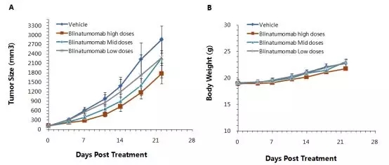
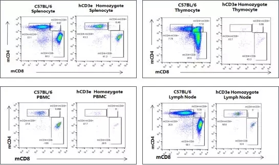
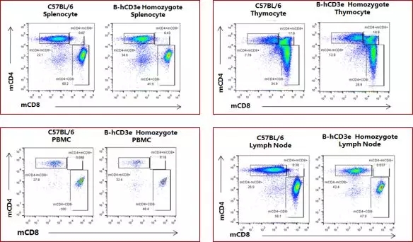
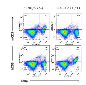
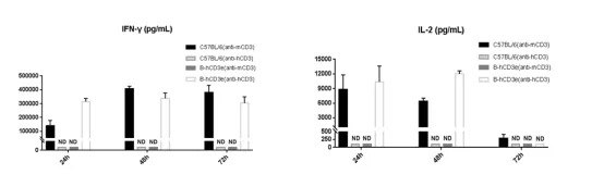
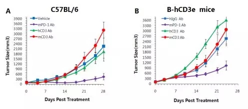
In this season of "grass long flying", bring you a new understanding of CD3e! CD3e (CD3e molecule) encodes a CD3-epsilon polypeptide that, together with CD3-gamma, -delta, -zeta and the somatic receptor alpha/beta or gamma/delta, constitutes a TCR-CD3 complex. This complex plays a very important role in antigen recognition and intracellular signal transduction pathways. In recent years, anti-tumor treatment using bispecific antibodies of human CD3 and tumor-associated antigen (TAA) has been extensively studied. However, due to the relatively low homology between the extracellular region of mouse CD3 complex and human species, human CD3-specific therapeutic antibodies cannot effectively activate mouse effector T cells, and an experiment suitable for evaluating human CD3-specific therapy is needed. Animal model. Humanoid CD3e mice (B-hCD3e mice) provide a powerful tool for the validation of double antibodies. TCR-CD3 complex and its signal pathway [1] mCD3e fully humanized mouse analysis The whole humanized mCD3e was replaced with hCD3e, and the spleen, thymus, PBMC and lymph nodes of the homozygous offspring were analyzed by flow cytometry to observe whether the T cell development of the mice was normal. The above results showed that compared with the wild type, hCD3e fully humanized homozygous mouse spleen, thymus, PBMC and lymph nodes had lower mCD3 + cells content and greater difference in CD4 and CD8 differentiation, indicating that the humanization strategy caused The development of T cells is blocked, and the mice prepared by this protocol are not suitable for subsequent pharmacological evaluation. Based on this, considering the influence of full humanization on the intracellular signal transmission of mCD3e, the Biotite map re-optimized the targeting scheme, only the mCD3e was humanized in the extracellular region, and the signal transduction function of the mouse source was retained to obtain B. -hCD3e mice. Flow cytometry analysis of spleen, thymus, PBMC and lymph nodes of homozygous offspring, the results are as follows: The results above show that, mCD3e extracellular domain of a humanized homozygous spleen, thymus, and lymph node of mouse PBMCs (B-hCD3e mice), the proportion of each cell and wild-type mice no significant difference, indicating that T cell differentiation Not affected by humanization of extracellular areas. Not everyone humanized mouse can have a normal function, not all CD3 humanized mouse called B-hCD3e mice Oh! Subsequent experimental data based on the in vitro detection and in vivo efficacy of the B-hCD3e mice model further confirmed the advantages of the model. The specific information is as follows: B-hCD3e mice basic information Basic Information Protein expression analysis Flow analysis showed: wild type C57BL / 6 mice was detected expression mCD3 + homozygous + B-hCD3e expression detected in mice hCD3. In vitro detection of CD3 antibody-activated T cells T cells (n=4) were isolated from spleen cells of wild-type C57BL/6 and homozygous B-hCD3e mice, and anti-CD3 antibodies (B-hCD3e, anti-hCD3; C57BL/6, anti-mCD3) and anti-CD3 antibodies mCD28 antibody incubation 24h, 48h, 72h. ELISA detected IFN-γ and IL-2. The results showed that: IFN-γ and IL-2 expression levels no significant differences in B-hCD3e and C57BL / 6. CD3 antibody efficacy test C57BL/6 (A) and B-hCD3e (B) were inoculated into MC38 cell line, and the tumor volume was about 150±50 mm 3 and then grouped (n=5). The results showed that mPD-1 antibody was significant in both mouse models. Inhibition of tumor growth indicated that humanization of extracellular domain had no significant effect on intracellular signal transduction of B-hCD3e mouse T cells. At the same time, hCD3e antibody significantly promoted tumor growth in B-hCD3e mice, but had no effect on tumor growth in C57BL/6 mice. Further studies showed that the AICD effect appeared after administration (hCD3e antibody), and T-cells of B-hCD3e mice were cleared, thereby promoting tumor growth. Flow cytometry to detect the ratio of B cells and T cells in peripheral blood The results showed that in the hCD3 antibody group, the proportion of T cells was significantly decreased due to the AICD effect caused by the hCD3e antibody. However, there was no significant change in the proportion of B cells. (A) There was no significant difference between the groups of CD19 cells compared to hIgG Ab. (B) A decrease in TCR-β% in the blood after h-CD3 Ab treatment compared to hIgG Ab. Bispecific antibody efficacy test based on B-hCD3e mouse The hCD19-MC38 cell line was inoculated subcutaneously into the mouse, and the tumor tissues were grouped (n=6) when they were close to 150±50 mm 3 , and different doses of Blinatumomab were injected at high, medium and low doses. The results showed that high doses of hCD3e bispecific antibody (Blinatumomab) significantly inhibited tumor growth. It was demonstrated that B-hCD3e mice can be evaluated for in vivo efficacy of bispecific antibodies. Tumor volume ± SEM B. A. Mouse mean body weight ± SEM. references [1]Immunology. 2018 Jan;153(1):42-50. doi: 10.1111/imm.12809. Epub 2017 Sep 5. List of Biographies anti-animal model List of Biographu double antibody cell line models Seafood Mix,Squid And Mussel Meat Mix,Monkfish And Shrimp Mix,Frozen Seafood Mix With Octopus Zhoushan Haiwang Seafood Co., Ltd. , https://www.haiwangseafoods.com
Background introduction 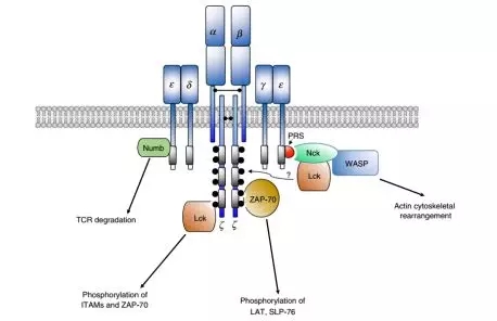

Shooting strategy 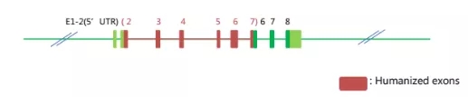
 Â