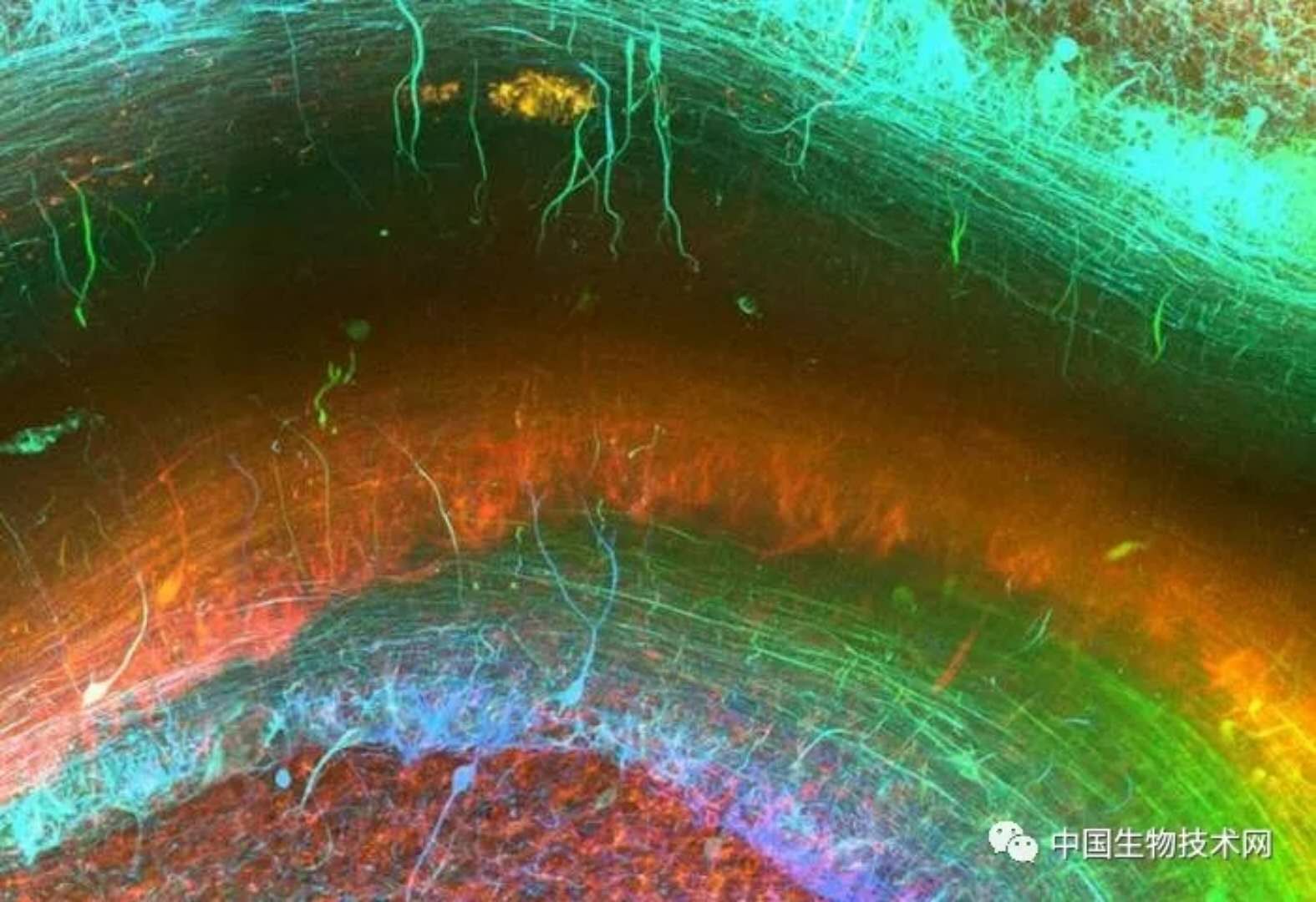
New technology "peeks into" the human brain from a three-dimensional perspective
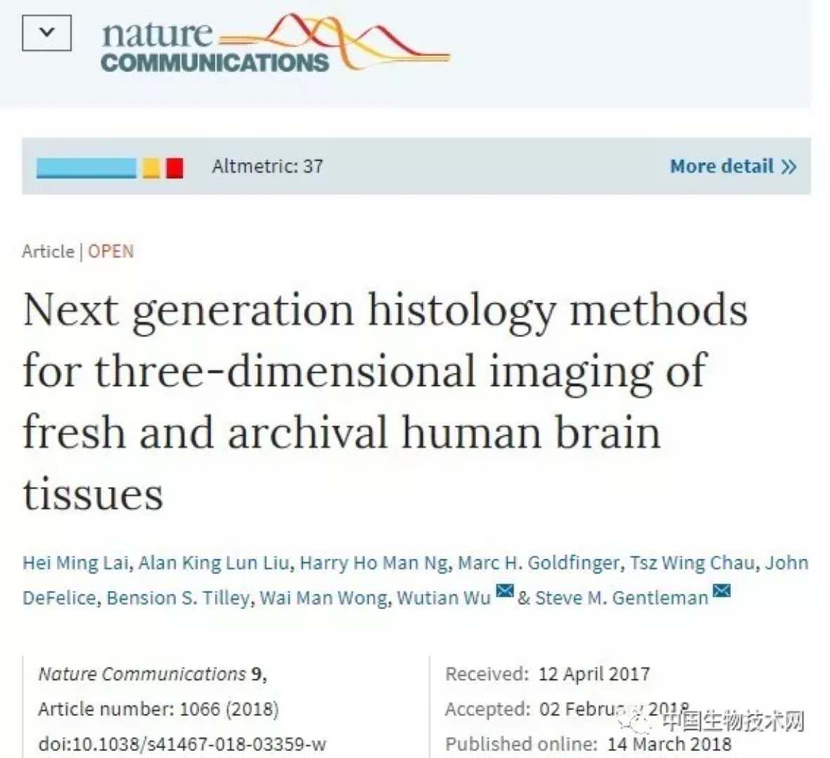
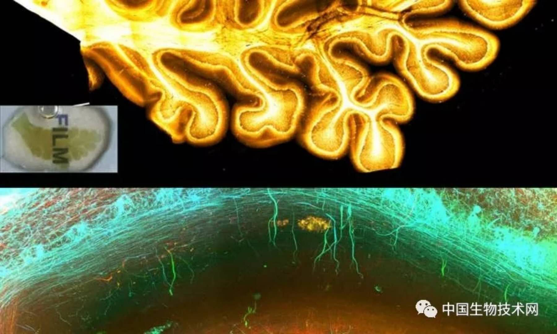
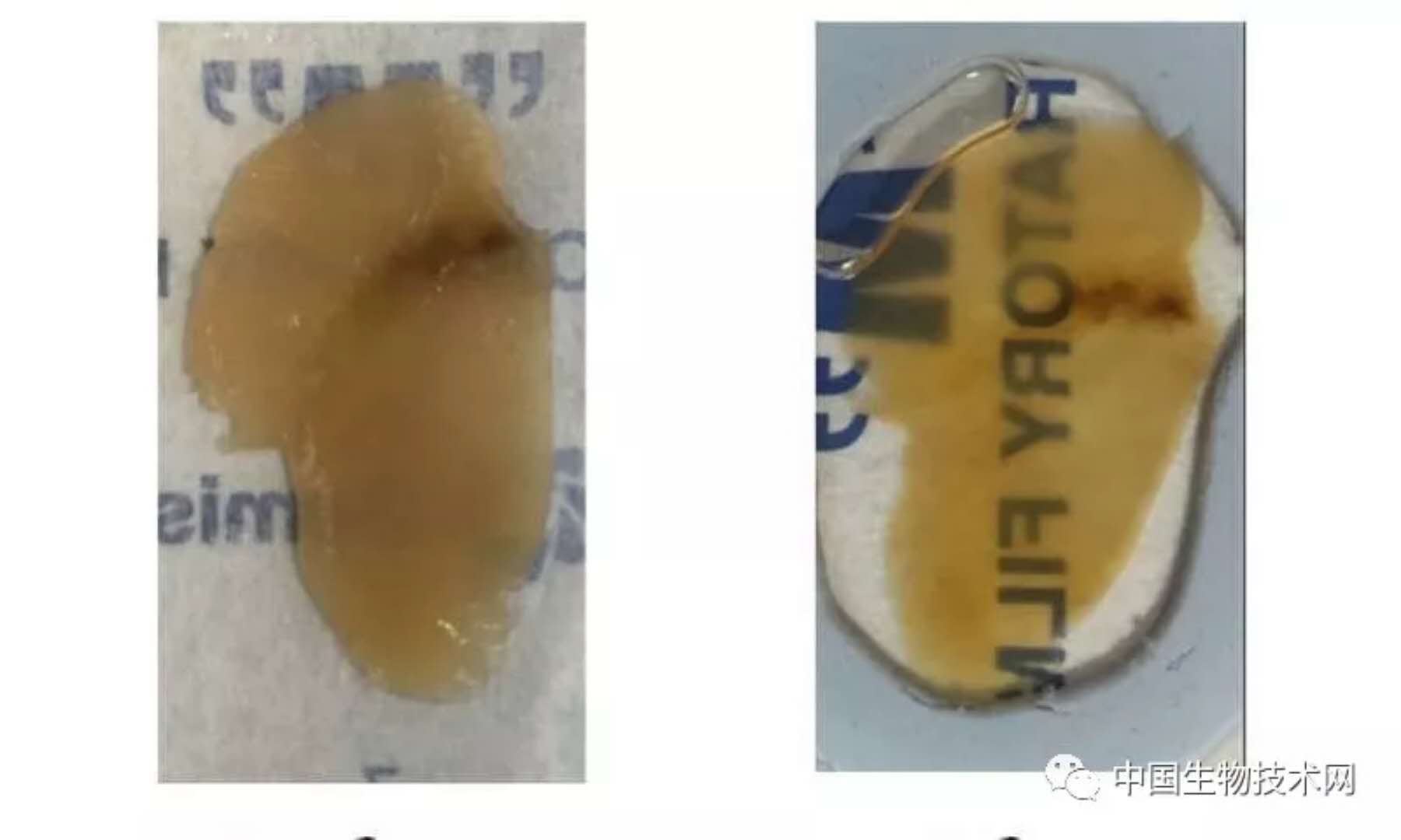
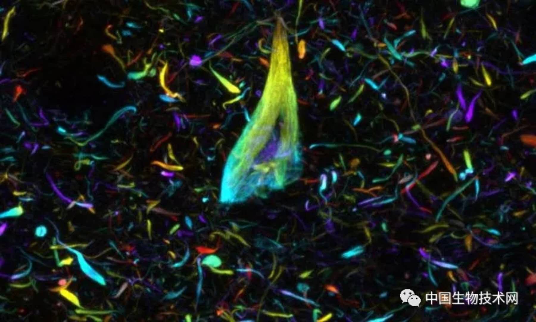
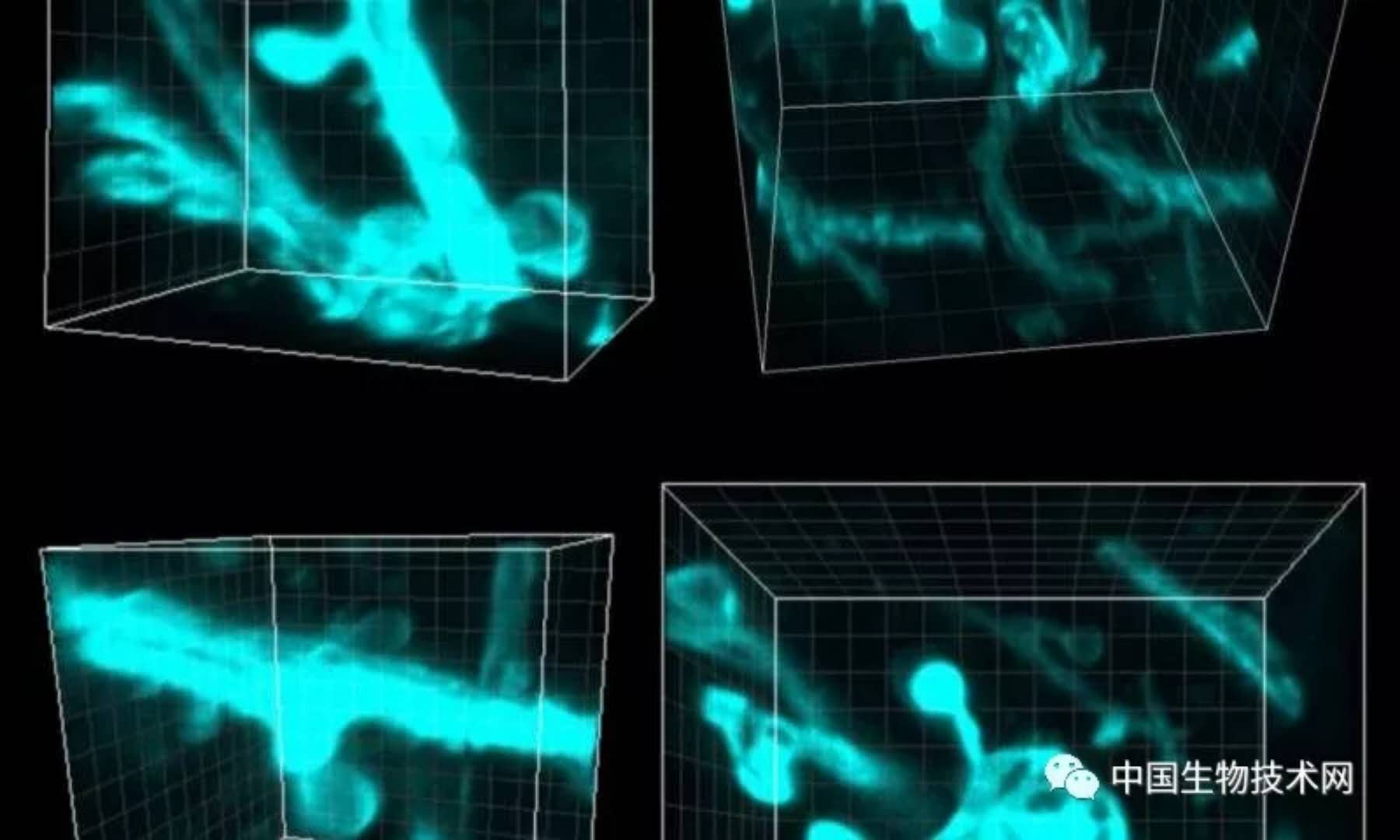
Release date: 2018-03-19 Amazing human brain tissue 3D version. Image source: Imperial College London Researchers at Imperial College of Science and Technology have developed a breakthrough imaging technique that reveals the extremely fine structure of the brain in unprecedented detail. The findings were recently published in Nature Communications. The next generation of technology allows researchers to generate 3D images of living brain tissue samples, which allows people to see amazing pictures of the human brain at the micro level. This achievement was done in collaboration with scientists from Imperial College and the University of Hong Kong. They believe that this technology can help explain the underlying mechanisms of many neurological diseases and bring the gospel to millions of people around the world. The traditional method of imaging brain tissue is to cut small pieces of brain tissue into ultra-thin sections and then color the sections to observe features of interest, such as proteins, or other disease-related biomarkers. However, in recent years, advances in molecular marker technology and the availability of laser microscopes have enabled researchers to develop modern tissue cleanup techniques. These techniques make the brain tissue transparent, allowing researchers to observe three-dimensional anatomy. However, these techniques were originally developed for brain tissue in rodents and are rarely used to study the human brain. Microscopic brain structure. Above: Cross section of the cerebellum. Below: A small internal image of the same sample showing the brain cell network. Image source: Imperial College London Many problems revolve around the unique nature of the human brain and the protection and processing of brain tissue after death. To overcome these problems, the research team led by three medical students developed a new tissue cleaning technique called OPTICLeal that allows multiple molecular marker technologies to be used for 3D visualization of living human brain tissue. Using this new method, they can stain nerve cells, glial cells, blood vessels, and other pathological markers, such as tau protein tangles in the brains of Alzheimer's patients. The details are extremely clear, and researchers can work in three dimensions. The middle connects them. Steve Gentleman, a scientist at the Imperial College of Parkinson's Brain Biobank, said: "These technologies allow us to reveal the microscopic structure of the human brain in astonishing detail." OPIClear technology enables researchers to “see through†the organization (left: before processing; right: after processing). Image source: Imperial College London He added: "By using such tools in the lab, we are able to visualize the three-dimensional processes of cell interactions and to better understand the pathways and connections that are impaired when neurodegenerative diseases affect a large number of patients. Thanks to the brain donors and their families, they made this research possible, and their selfless spirit deserves our respect." Tau protein tangles in the brain of Alzheimer's patients. Image source: Imperial College London According to the researchers, the method is relatively inexpensive, time efficient and easy to implement, and is likely to be the basis for future technological development. They hope that a better understanding of the connections and circuits in the brain at the micro level will help us gain insight into the pathology of common neurodegenerative brain diseases such as Alzheimer's and Parkinson's. A dendritic spine, a protrusion of the axon tail of a brain cell that allows signals to pass through the cell. Image source: Imperial College London Source: China Biotechnology Network Pacific Mackerel Pacific Mackerel ZHEJIANG EVERNEW SEAFOOD CO.,LTD , https://www.evernewseafood.com