Application of Feina desktop scanning electron microscope in lithium battery separator industry
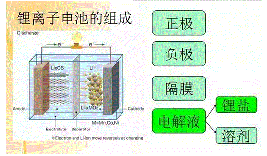
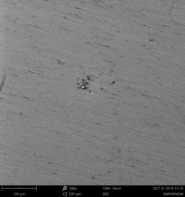
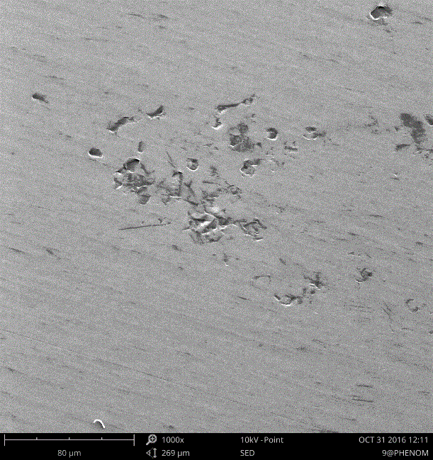
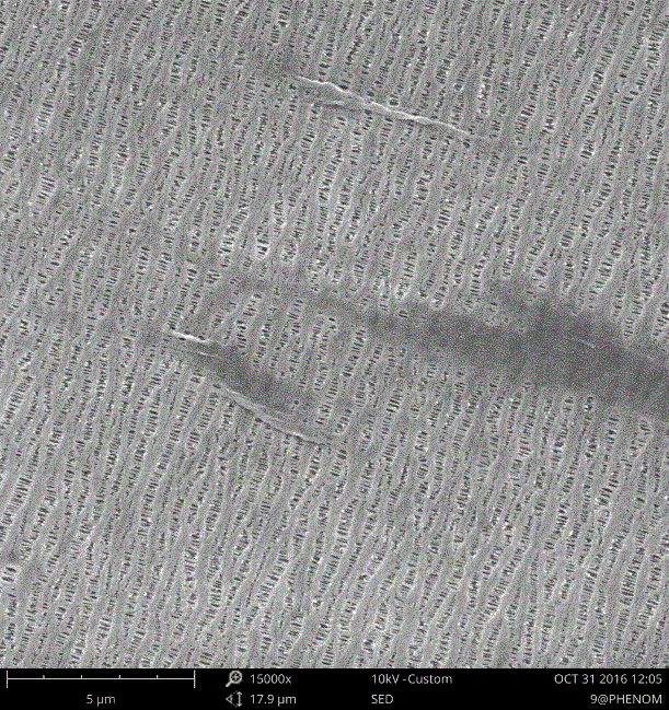
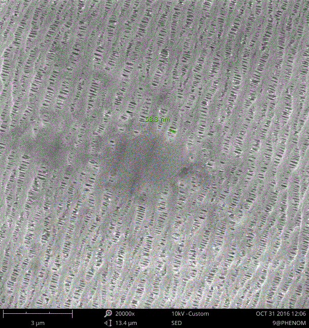
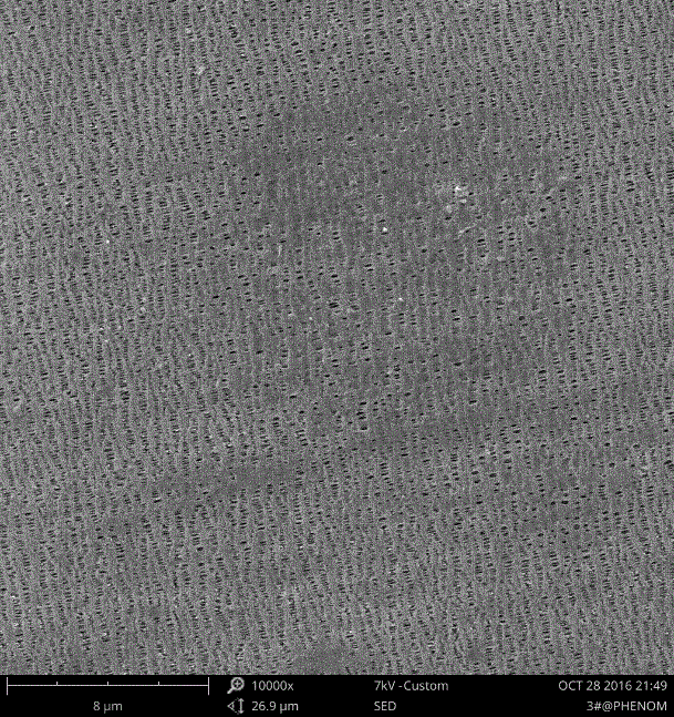
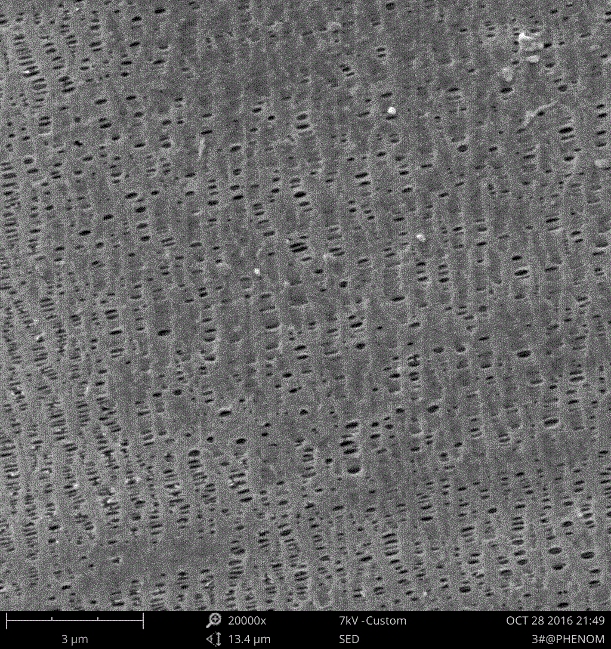
In the structure of the lithium battery (see Figure 1 below for the structure of the lithium battery), the diaphragm is one of the key inner components. The performance of the diaphragm determines the interface structure and internal resistance of the battery, which directly affects the capacity, circulation and safety of the battery. The separator with excellent performance plays an important role in improving the overall performance of the battery. The evaluation of the diaphragm performance needs to be detected by scanning electron microscopy. Especially for the lithium battery series, since the electrolytic solution is an organic solvent system, it is also required to use an organic solvent-resistant separator material, and a high-strength thinned polyolefin porous film is generally used at present. Figure 1. Schematic diagram of the structure of a lithium battery In order to ensure low resistance and high ionic conductivity, lithium ion is well permeable, and the diaphragm must have a certain pore size and porosity. In order to verify the diaphragm's ability, scanning electron microscopy is required. Microscopic observation ensures that the pore size and size range of the membrane and the pore diameter are uniform, and there are defects such as scratches and pits on the membrane. To this end, the Fein desktop SEM observed the battery separator. The following images are part of the results: Figure 2 The left and right images are taken at low magnification with a Fein desktop SEM. You can see some pit defects on the film and some uneven stretching. Figure 2 Feina desktop scanning electron microscope low magnification diaphragm test results Left: 500 times result graph right: 1000 times result graph Figure 3 The left and right images are also the results of the observation of the film by the Feiner desktop scanning electron microscope. The left picture is a photo enlarged to 15000 times. The surface of the film has a large number of apertures. The right picture shows one of the holes. The size is 58.3nm; the pore size of the membrane is between 10 and 100nm, which is relatively uniform, but there is no stretching in some places, which will affect the performance. Figure 3 Feina desktop scanning electron microscope observation of the film Left: 15000 times result graph right: 20000 times result graph Figure 4 The left and right figures are another defect on the diaphragm observed by the Feiner desktop SEM. The aperture is slightly larger than normal. Figure 3 Another defect map observed by the Fein desktop SEM Left: 10000 times result graph right: 20000 times result graph Baby Hair Clippers,Hair Trimmer,Hair Cutting NINGBO YOUHE MOTHER&BABY PRODUCTS CO.,LTD , https://www.oembreastpump.com