Application of filters on confocal microscopy
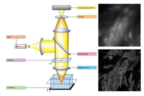
Due to the unique arrangement of the apertures, the images provided by the confocal microscope detector correspond to a thin optical section or a section of the sample. For example, a sample that is a few millimeters thick is reduced to less than a micron along the z-axis at the focal plane. The illumination field at the confocal point is limited by an aperture called a pinhole. The field of view is also limited by a pinhole that is positioned relative to the image plane conjugated to the first aperture and the illumination point. The result of this confocal configuration is a reduction in the detection of defocused light, thereby increasing the signal to noise ratio by reducing the imaging volume. Marvin Minsky first designed a confocal microscope and described it in a 1957 patent by moving the beam platform. The modulated light from the sample is sent to the photocell for observation with an oscilloscope. Light illumination and subsequent detection are achieved by moving the platform through the point of view. Minsky's original conception completely changed the microscope. When Minsky's patent was passed, there were still several technical difficulties. His system requires a very strong light source due to the limited aperture. In view of the light intensity lost by the pinhole, the laser did not exist at the time, and there were no other sufficiently strong light sources to excite the fluorescence. At that time Minsky used a long-lasting oscilloscope to make observations, so there was no way to record the images faithfully. It has led other inventors to further develop this special microscope as an effective tool for ordinary researchers. The development of a strong monochromatic light source, the laser, solves the problem of illumination. The computer is equipped with a digitizer to make recording and realistic images a breeze. Today, scanning confocal uses a laser or a combined laser as an illumination source. This scanning is done by precisely controlling the galvanometer in a grating motion, sweeping the field of view with a minimum spot, just like controlling a TV through a computer. The fluorescent signal scattered or reflected from the sample is sent to a photomultiplier tube (PMT), which is a point (like an element, a pixel point) that forms an image on the screen at a time. Although Minsky lists many of the advantages of confocal, perhaps the most important feature is the scanning confocal microscope that enables optical section imaging of samples. Traditionally, the fine structure of a cell or tissue is obtained by fixing the tissue and then carefully slicing it into a thin layer for observation and imaging. This process requires the sample to be sacrificed and requires the researcher to spend several years learning the slicing technique to cut the sample to a sufficiently thin thickness for imaging. Confocal optical slice performance allows users to image thick tissue without the need for special slicing techniques. It also allows users to perform ultra-high resolution imaging of living cells, tissues and organisms. Living cell imaging has become an important part of confocal microscopy. This ability to slice optically also means that a single slice/image can be saved to a computer and then reconstructed into a three-dimensional image of the sample. This is a very important feature in existing laser scanning systems. Sunflower Seeds,Sunflower Kernels,Raw Sunflower Seeds,Bulk Sunflower Seeds Inner Mongolia Hengxintonghui Supply Chain Management Services Co.,LTD , https://www.hxthfood.com
This system uses some special optical components in fluorescence imaging. A common misconception is that the laser produces a laser output with only one wavelength. Virtually all lasers will actually generate harmonics or scatter other wavelengths of light. Although the light in these sub-bands is weak compared to the main band, they can still greatly reduce the signal-to-noise ratio. If the emission band of the fluorescence falls within the range of the laser harmonics (or other stray light from the laser), the fluorescent signal may be completely masked and undetectable.
The primary device in the optical path is the laser purification filter, which is the modified excitation light filter. Due to the coherence of the laser and the relatively small beam and alignment of the optical path, these lenses are ground and polished. This is in stark contrast to wide field microscopes because wide field microscopes do not require grinding and polishing. Purified lenses should also have good transmission characteristics, including wavefront distortion of less than one wavelength per inch. The off-angle (deviation from the ideal parallel line to the outer edge of the lens) should be reduced to less than one arc angle, so that it is not necessary to re-correct when using different purification filters in the same system.
The half-wave width (FWHM) of a purification filter is usually around 10 nm. It blocks other stray light from the laser source (maximum range from UV light to 1200 nm) and anti-reflective coating for maximum transmission.
With the advent of newer, more powerful lasers, it is not necessary to use an anti-reflection coating to increase transmission. However, these optics use a maximum reflective interference coating to avoid thermal damage, which will therefore reduce surface reflection. These reflected light cannot be reflected into the resonant cavity of the laser. Therefore, these light pieces are designed for an incident angle (AOI) between 3.5 and 5 degrees. The wavelength of the light emitted by the diode laser after warming up will vary slightly. Therefore, we recommend that these sources be equipped with a 20-25 nm wide purification filter. In confocal systems and common fluorescence microscopy systems, in order to achieve better planar flatness, they must overcome wavefront aberrations. Although the wavefront aberrations of both systems are controlled within a range of one wave per inch, it is clear that a dichroic mirror can be selected for better results in a confocal system.
At present, it is common to achieve dichroism on a 4-6 m thick fused silica substrate, but more and more demands require thicker substrates to eliminate wavefront aberrations and achieve dichroism.
Over the past few years, as lasers have grown in size, there will be more and more power loads that could ruin the main mirror. This is not a problem for a well-designed dichroic mirror with a beam load of at least 8-10 watts for a common beam size. The transmission is maximized by the AR coating to minimize the reflection of the focused light.
Polarization is another factor to consider in the filter. Since all of the light members can act as polarizers when they have a deflection angle to the optical path, this is especially important for most polarized lasers.
The primary role of all fluorescent emission filters is to prevent the transmission of excitation light. Compared to fluorescence microscopy, for confocal systems, this blockage does not encompass a wide spectral range. However, due to the high power output of the laser beam, it is necessary to specifically design a larger blockage (perhaps above OD 8) for the specific wavelength of the laser emission.
There is still some controversy about the actual design of the confocal system emission filter. Since most detectors use a photomultiplier tube (PMT), the image captures only one pixel (image element), and these filter components do not need to be polished and polished. However, because confocal imaging has higher and higher resolution requirements, it is not a waste of effort to polish and polish like other transmitting filters. Recent evidence suggests that when the transmission filter has a large wedge shape (beam degradation) may cause inaccuracies in morphological measurements, which may be more severe in long-emitting optical paths.
The filter optics housing of the laser system is designed around the laser emission and does not necessarily target the maximum or excitation of a particular fluorescent dye. This does bring a few questions about what kind of fluorescent dyes are suitable for a particular laser system. Fortunately, there are now more options available from suppliers of fluorescent dyes. Alternatively, you can choose to use a multi-color primary mirror that can easily and quickly reflect multiple laser beams.